




If you’ve ever wondered, “what is a clinical breast exam?”, here is the short answer. A clinical breast examination is a hands-on check of your breasts, chest wall, collarbone area, and underarms, performed by a trained healthcare provider such as a primary care physician, OB/GYN, or nurse practitioner. Your provider looks for lumps, skin changes, nipple changes, and swollen lymph nodes.
A clinical exam is different from a self-check at home. A breast exam by a doctor uses a systematic technique and a trained touch to notice subtle differences in tissue. It usually takes just 5 to 10 minutes and needs no special equipment. It is often part of a routine well-woman visit and one piece of broader breast cancer screenings.
Knowing what to expect takes much of the nervousness out of the appointment, whether it’s a routine preventive visit or a follow-up after breast cancer treatment.
Why Do Doctors Perform Breast Examinations?
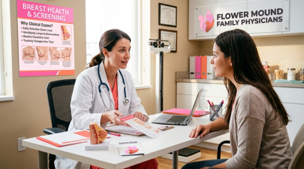
Doctors perform breast examinations to learn what is normal for your body and to catch changes that deserve a closer look. Breast tissue changes with age, hormones, pregnancy, and your menstrual cycle, so a provider who sees you regularly is well placed to notice what’s new.
The importance of clinical breast exams for women’s health shows up in several situations:
- Evaluating new symptoms. A new lump, localized pain, skin dimpling, or nipple discharge is usually assessed by a physician first. The exam helps decide which imaging test, if any, comes next.
- Adding to mammography. Mammograms are the most effective tool for finding tumors too small to feel, but no test catches everything. Dense breast tissue can hide small abnormalities, so a physical exam adds another layer of assessment.
- Monitoring higher-risk patients. For breast cancer survivors and women with a genetic mutation such as BRCA1 or BRCA2, exams help track the chest wall, surgical scars, and lymph nodes between scans.
- Building breast awareness. Your provider can show you what normal tissue feels like, including ridges and natural lumpiness, so you can recognize changes sooner.
What Happens During a Clinical Breast Examination?
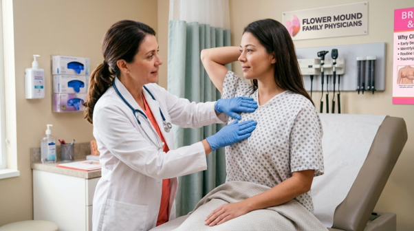
Here is what to expect during a clinical breast exam with a doctor. Details vary by provider, but most visits follow the same four steps.
1. Health history and symptom review
Before the physical portion, your provider will ask about your menstrual cycle, pregnancies, hormone therapy, previous biopsies, family history, and any changes you’ve noticed. If you’ve had breast cancer, they’ll also review your surgery, radiation, and current surveillance plan.
2. Visual inspection
You’ll undress from the waist up and wear a gown that opens in front. A chaperone may be present, which is standard practice. While you sit upright, your provider looks for:
- Differences in size, shape, or contour between the breasts
- Skin dimpling, puckering, or thickening, sometimes called peau d’orange
- Redness, scaling, or a persistent rash on the nipple or areola
- A newly inverted nipple or discharge
You may be asked to raise your arms, press your hands on your hips, or lean forward. These movements tense the chest muscles and make subtle skin changes easier to see.
3. Palpation while lying down
Next, you’ll lie back with one arm raised over your head. This spreads the breast tissue thinly across the chest so it’s easier to examine. Using the flat pads of the middle three fingers, your provider works through the whole breast in an overlapping pattern, such as vertical strips, and applies light, medium, and firm pressure at each spot to reach different tissue depths.
The exam covers the entire breast area, from the collarbone down to the bra line and from the breastbone out to the middle of the underarm.
4. Lymph node and nipple check
Because lymph fluid from the breast drains toward the underarm, your provider will check the axillary lymph nodes and the areas above and below the collarbone. Healthy nodes are soft and hard to feel. Nodes that are firm, enlarged, or fixed in place need follow-up. Finally, the provider gently checks the nipple and areola for tenderness or discharge.
Is a Clinical Breast Exam Painful?
A clinical breast exam should not be painful. You may feel mild, brief pressure when your provider examines deeper tissue near the ribs. If your breasts are tender before your period, or if you have scar sensitivity after surgery, tell your provider at the start so they can adjust their pressure. You can also ask them to pause at any point.
Clinical Breast Exam vs. Mammogram vs. Self-Exam
Each method plays a different role in breast health and early detection.
Method | Who performs it | Purpose | Main limitation |
Clinical breast exam | Physician, OB/GYN, or nurse practitioner | Hands-on and visual check of breast tissue and lymph nodes | Can’t detect microcalcifications or very small, deep tumors |
Screening mammogram | Radiologic technologist; read by a radiologist | Low-dose X-ray of internal breast tissue | Dense tissue can sometimes hide small lesions |
Breast self-awareness | You, at home | Knowing your normal so you notice changes | Not a substitute for professional care; can cause false alarms |
What is the Difference Between a Clinical Breast Exam and a Mammogram?
A clinical breast exam relies on a provider’s touch and eyes to find palpable lumps, skin changes, or swollen lymph nodes. A mammogram uses low-dose X-rays to create images of the inside of the breast, and it can spot tiny tumors, sometimes years before they can be felt. The two work best together. A mammogram sees what hands can’t, and an exam can notice surface, chest wall, or underarm changes that imaging may not capture well.
Can a Clinical Breast Exam Detect Breast Cancer?
A clinical breast exam can detect physical signs that may point to breast cancer, such as a firm, painless lump, skin dimpling, or enlarged underarm lymph nodes. It cannot diagnose cancer on its own. Only imaging and a tissue biopsy can confirm what a finding is.
Most lumps found during an exam turn out to be benign, such as fluid-filled cysts, fibroadenomas, or normal hormonal lumpiness. Still, any new lump deserves evaluation.
When to Schedule a Routine Breast Examination
When to schedule a routine breast examination depends on your age, history, and risk factors. Guidelines differ between organizations, and some do not recommend routine exams for average-risk women, so it’s worth discussing what is right for you:
- Ages 20 to 39, average risk: Many providers offer a clinical breast exam every 1 to 3 years as part of a well-woman visit. ACOG’s guidance for average-risk women begins around age 25.
- Age 40 and older, average risk: Providers commonly offer an exam during annual wellness visits alongside regular screening mammograms.
- Higher risk and survivors: If you have a personal history of breast cancer, atypical hyperplasia, chest radiation before age 30, or a known genetic mutation, your care team may recommend exams every 6 to 12 months along with mammography and sometimes breast MRI.
- Any age with new symptoms: Don’t wait for your annual visit if you notice a new lump, ongoing focal pain, skin changes, or bloody nipple discharge.
At what age should women start breast cancer screening?
Women at average risk should begin screening mammograms at age 40. The U.S. Preventive Services Task Force recommends mammography every two years from ages 40 to 74. The American Cancer Society says women can choose to start at 40, screen yearly from 45 to 54, and then move to every two years or continue yearly from 55. Women with a strong family history or genetic risk may need to start earlier, so ask your doctor about a personalized plan.
How Doctors Evaluate Unusual Breast Changes
When a patient has a concern, or an exam turns up something new, doctors follow a step-by-step process instead of relying on touch alone. Here is how doctors evaluate unusual breast changes.
What they assess during the exam
- Size and shape. Smooth, round, well-defined borders often suggest a cyst or fibroadenoma. Irregular or poorly defined edges call for closer imaging.
- Benign lumps often move easily under the skin, while suspicious ones may feel fixed to surrounding tissue.
- Soft or rubbery areas feel different from hard, dense nodules.
- Your provider compares the same spot on the other breast to see whether it’s normal anatomy.
Possible next steps
- Diagnostic mammogram: Magnified, angled views of the area of concern, unlike a routine screening mammogram.
- Targeted breast ultrasound: Sound waves show whether a lump is a harmless cyst or a solid mass, which is especially useful in dense breasts and in women under 30.
- Core needle biopsy: If imaging is unclear or suspicious, a radiologist takes a small tissue sample under local anesthesia, and a pathologist examines it for cancer cells.
How to Prepare for Your Appointment
- Pick your timing. If you still have periods, try to book 7 to 10 days after your period starts, when breasts are usually least tender.
- Skip deodorant, powder, and lotion if a same-day mammogram is possible. Some products can appear as white spots on images and mimic calcifications.
- Wear a two-piece outfit so you only need to undress from the waist up.
- Bring records and questions, especially prior mammogram reports, ultrasounds, or pathology results if you’re seeing a new provider.